Wednesday, February 21, 2024
By Jenny Blair, images by Jon Atherton
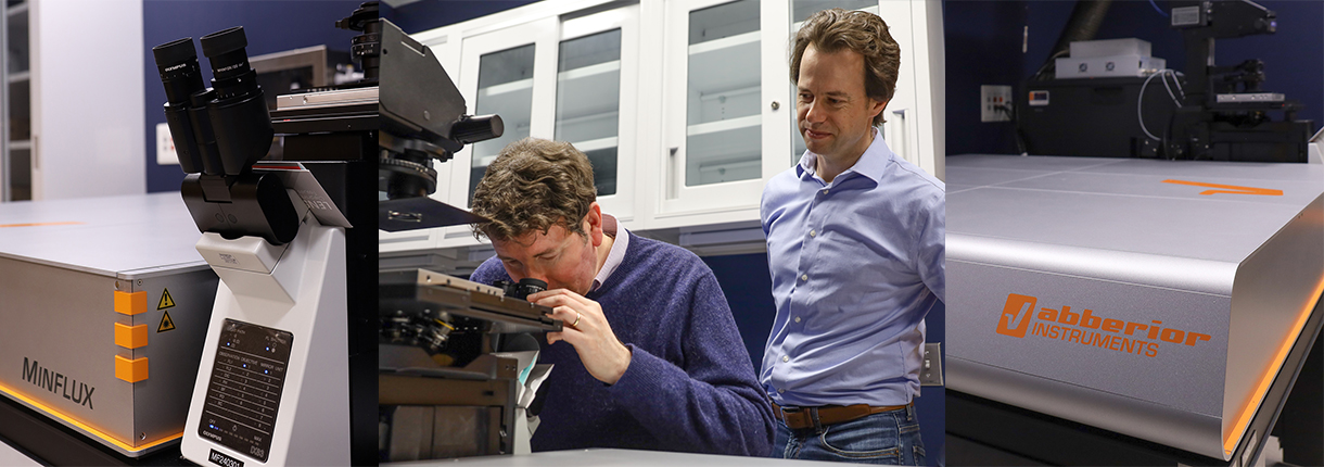
Yale researchers will soon watch individual molecules move through living cells, thanks to the arrival of an Abberior MINFLUX instrument, the world’s most powerful 3D super-resolution fluorescence microscope.
Coupling 2- to 3-nanometer resolution in three dimensions with speeds that are orders of magnitude faster than camera-based systems, the MINFLUX at Yale is one of only four such microscopes in the country and the first to be installed in the entire Northeast.
“For fluorescent microscopy, this level of resolution in 3D has not been done before—not anywhere close,” says Joerg Nikolaus, director of the West Campus Imaging Core, where the microscope will be housed. The core is one of multiple shared imaging facilities at Yale.
By comparison, super-resolution microscopy achieves 20- to 30-nanometer resolution, while confocal microscopy has an approximately 200- to 300-nanometer capability.
The new microscope’s molecule-scale resolution in not only the xy plane but also the z-axis means it can reveal whether two molecules are close enough to physically interact.
John D. MacMicking, PhD, a Professor of Microbial Pathogenesis and of Immunobiology and a Howard Hughes Medical Institute (HHMI) investigator, purchased the instrument thanks to generous support from HHMI, which supports its investigators to pursue important biomedical questions. From his lab at the Yale Systems Biology Institute, MacMicking studies how cells defend against infection.
The objects he deals with–viruses and bacteria, for example–are miniscule in size, so the MINFLUX microscope is the ideal tool to investigate cellular events critical for controlling human pathogens.
Using hollow sphere or “bottle-beam” laser excitation technology, the new microscope allows researchers to determine protein stoichiometries, or how many of each protein type make up a given protein complex. It uses fewer photons, which means less phototoxicity.
That allows for longer capture times and the ability to see proteins’ locations in three-dimensional space, MacMicking explained.
“If you’ve got sufficient photons to know where a protein is localized, you can track that individual protein and follow the kinetics of what it is doing in the cell—observed at speeds and with a precision that you couldn’t do using conventional cameras,” he said. “It allows us to start to bridge light microscopy with electron microscopy.”
MacMicking plans to use the instrument to build upon forthcoming published research in which his lab used CryoEM and CryoET to visualize antimicrobial proteins in action against bacteria and viruses, including those immune proteins which inhibit SARS-CoV-2 infection.
“We can study them in real time under physiologically relevant conditions to determine how they’re resolving the infection, whether they bring other host protein partners with them, and what those partners do,” MacMicking said. “You start to get functional high-resolution imaging.”
Find out more about Yale’s research core technology at research.yale.edu.